Disarmed A. tumefaciens strain (e.g. EHA101) harboring binary vector of interest
For
this protocol it is assumed that the vector harbors the nptII selectable marker gene and the uidA scorable marker gene (e.g. pDE00.0201 from Reference 2).
Chemicals
Driver Kuniyuki walnut (DKW) medium with vitamins (PhytoTechnology Laboratories®, catalog number: D2470)
Top loading electronic balance (e.g. Mettler, model: PM 2000)
pH meter (e.g. Corning Pinnacle, model: 540)
Constant temperature incubator (e.g. Napco, model: 301)
Water baths (e.g.Thermo Scientific, model: 2870)
Vortex mixer (e.g.: Scientific Industries, model: G 560)
Freezer (- 80 °C) (e.g. New Brunswick, model: U 700 Premium)
Procedure
Obtain actively multiplying walnut somatic embryo cultures or initiate new cultures from zygotic embryos or anther tissue (Dandekar et al., 1989; Mendum and McGranahan, 1995; Leslie et al., 2006).
Several days before initiation of transformation:
Streak Agrobacterium strains out on a LB plates with appropriate antibiotics. Incubate at 28 °C for 48 h.
Culture sufficient numbers of embryos of the desired genotypes.
Prepare appropriate antibiotics.
Make LB liquid medium.
Make co-cultivation liquid medium.
Make DKW basal, AS, and selection media.
The day before initiation of transformation:
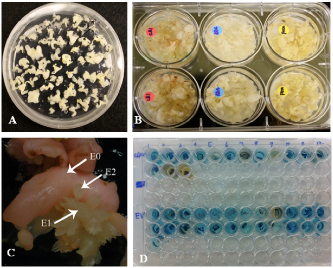
Select embryos: Pick out 60-70 or more rapidly growing, small, white, uniform embryos (Figure 1A) and place them onto fresh DKW basal medium.
Day 1 (prepare liquid cultures)
Inoculate each strain of Agrobacterium to be used into a 50 ml conical centrifuge tube containing 20 ml of LB medium.
Place the capped tubes on a rotary shaker at ~200 rpm at 25 °C.
After 2 h add the appropriate selective antibiotics for the vector used and return to shaker.
Day 2
After shaking overnight, the bacterial cultures should be turbid. Determine the Abs600nm of a 10-1 dilution of the culture.
For 60-70 embryos, approximately 25 ml of co-cultivation suspension
will be utilized. The desired Abs600nm of the co-cultivation suspension
is ~0.5 (an Abs600nm reading of 0.5 is approximately equivalent to 2.5 x
108 bacteria/ml). To calculate the needed volume of overnight culture,
use the following equation:
(Co-cultivation volume needed) x (desired Abs600nm of the co-cultivation suspension)
(Abs600nm of the Agrobacterium culture) x (dilution factor)
For
example: If you need 25 ml of co-cultivation suspension at a
concentration of 2.5 x 10⁸ bacteria/ml and an aliquot of the Agrobacterium culture diluted 1: 10 gives an A600 reading of 0.371, how
much of the Agrobacterium culture do you need to dilute for use?
(25) x (0.5) = 3.36 ml
(0.371) x (10)
Using a sterile pipette, place the calculated volume of Agrobacterium
culture into a sterile plastic 50 ml centrifuge tube and centrifuge for
10 min at 4,000 x g and ambient temperature to pellet the bacteria.
Pour or pipette the supernatant into a waste container and resuspend
the pellet in the co-cultivation medium. The pellet is easier to
resuspend in a small volume (0.5 ml first) by carefully pipetting up and
down, followed by bringing the solution to required final volume.
Return the tubes to the rotary shaker at ~200 rpm at 25 °C for 1-2 h.
Co-cultivation
Place desired number of embryos into a well of a sterile six-well
multiwell plate. The well should be no more than half filled with
embryos (about 20-23 embryos). Use more than one well for each
transformation in case of contamination problems. Transformations of
multiple genotypes can be performed in different wells of the same plate
but if using multiple bacterial vectors it is advisable to use separate
plates to avoid cross-contamination.
Dispense the
appropriate volume of Agrobacterium co-cultivation suspension (about 8
ml, or enough to cover the embryos) into each well using sterile 10 ml
pipettes with cotton-plugged ends.
Incubate at ambient temperature for at least 10-15 min (Figure 1B and Note 1).
Place a sterile filter paper into a number of empty sterile Petri plates equal to the number of wells used.
Pipet as much excess co-cultivation liquid as possible from each well into a waste container.
Transfer the embryos from each well onto the filter paper in the Petri
plates using sterile forceps. This will wick excess co-cultivation
liquid from the embryos.
Transfer the embryos to labeled plates
of AS medium (about 10 per plate) and place the plates in the dark for
48 h at 20-22 °C (see Video 1).
Video 1. Walnut transformation
Day 4: Selection
After co-cultivating for 48 h, transfer the embryos to plates of KAN/Timentin medium containing 200 mg/L kanamycin and 200 mg/L Timentin (see Note 2). Other selection medium may be utilized depending on the selectable marker gene (see Note 3). Incubate the culture plates in the dark at ambient temperature (20-24 °C).
Day 6 and onward:
Transfer embryos to fresh KAN/Timentin medium after two days and again
after an additional five days. This helps to reduce bacterial
overgrowth. Thereafter transfer the embryos to fresh KAN/Timentin medium
weekly for 8-12 weeks.
As new somatic embryos begin to develop
from the surface of the original (E0) embryos, separate them from the
parent embryos. Label these new embryos as E1 embryos. Repeat this
process for one more generation (E2 embryos; Figure 1C).
After
8-12 weeks of selection, embryos can be moved to selection medium
containing only kanamycin. Observe the embryo cultures carefully for the
next several weeks to ensure that no residual Agrobacterium has
survived.
Scoring for GUS expression
As E2 embryos emerge, test them for GUS (uidA) activity (Jefferson, 1987).
Pipette 40 µl of X-gluc staining solution (see recipes) into wells of a sterile 96-well multiwell plate.
Using a fine point scalpel remove a small piece of tissue (cotyledon
tips work well) from each well-formed and healthy E2 embryo of interest.
Put the tissue piece in the X-gluc solution and label and mark the
location of the embryo from which it was excised.
Incubate at 37 °C and monitor the development of blue color. Color change should be apparent in 10 min to 2 h (Figure 1D).
If the tissue piece developed the distinctive blue color, the E2 embryo from which it was cut should be separated and multiplied on
selection medium until E3 embryos are available for DNA analysis.
Figure
1. Walnut transformation. A. Embryos with well-develped cotyledens
ready for transformation. B. Embyos in co-cultivation suspension. C.
Primary embryo (E0) developing secondary embryos (E1 and E2). D. GUS
test, transformed embryos turn blue in X-Gluc solution.
PCR based verification of transformation
Select actively proliferating E3 embryos from different embryo lines and use for DNA isolation.
Isolate total DNA using a DNeasy Plant Mini Kit according to the manufacturer’s protocols.
Perform PCRs using appropriate primers to confirm the presence of the
nptII gene (or other gene of interest). Set up the PCR reactions using
250 ng of genomic DNA in each 25 μl PCR reaction.
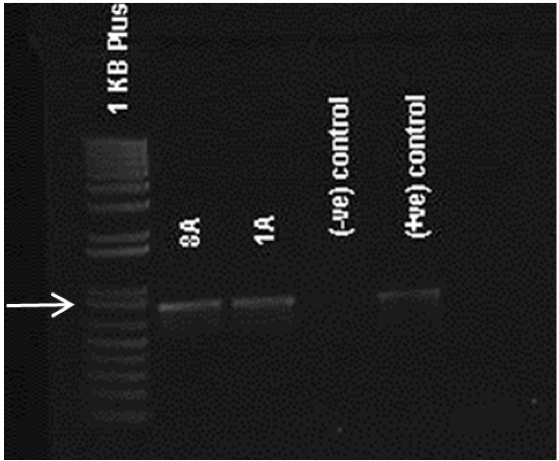
After PCR is
completed, 5 μl of PCR product is electrophoresed to verify
amplification of an appropriately-sized band. DNA from untransformed
embryos can be used as a negative control (Figure 2).
Figure 2.
Confirmation of transgenic nature. Agarose gel of PCR products showing
790 bp bands in transformed lines 8A and 1A for APh3/Aph4 primers.
Negative controls or non-transformed lines does not show any bands.
Somatic embryo germination and plant production
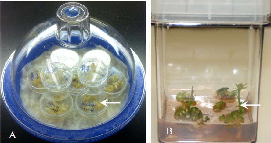
After verification of transformation, some E3 embryos from the desired
embryo lines can be desiccated to initiate germination. Choose
well-formed somatic embryos and place them in 35 x 10 mm sterile Petri
plates with no medium. Cover the plates but leave them unsealed (do not
wrap with Parafilm®) and place them in the dark at ambient temperature
(20-24 °C) on the rack of a well-sealed desiccator containing 10-15 ml
of saturated ZnSO4 or NH4NO3 in the bottom (Figure 3A).
After
the embryos become an opaque white (typically 2-7 days) remove the
embryos from the desiccator and place them on DKW shoot medium in
Magenta GA-7 vessels or Petri plates.
Culture at ambient temperature under cool white fluorescent lights (16 h day length, ~100 µE) for 2 to 8 weeks.
Most embryos will produce roots, but typically fewer than 10% of
embryos develop shoots. Roots will usually emerge from embryos in 7-10
days (Figure 3B).
Fully germinated embryos possessing both
roots and shoots should be removed from the medium as soon as possible
and planted in potting soil (for example UC Mix, 25%: 42%: 33% sand: fir
bark: peat moss). Alternatively, epicotyls can be excised,
micropropagated on DKW shoot medium, and then rooted to generate
multiple plants.
To acclimatize, keep plants on soil at 100% humidity for 2 weeks and then gradually reduce the humidity.
Established plants can be repotted to larger containers as needed and maintained in a greenhouse or lath house.
Figure
3. Plant production using trnasgenic somatic embryos. A. Drying embryos
in dessicator B. New shoots emerging from transformed walnut somatic
embryos.
Notes
Physical wounding is not necessary when somatic embryos are used for transformation.
Transfer embryos in a well-spaced pattern on each plate so that if Timentin-resistant bacteria begin to multiply they are not moved to all the embryos on the plate.
Hygromycin B (25 mg/L) can serve as an alternative selectable marker in place of kanamycin or as a second selectable marker if performing co-transformation to insert two genes simultaneously.
Kanamycin sulfate in solution has a very high pH. If used at a concentration greater than 100 mg/L for selection adjust the pH of the kanamycin stock solution to 5.5 with dilute HCl prior to filter sterilizing.
Recipes
Kanamycin sulfate (50 mg/ml stock)
5 g dissolved in 100 ml dH2O
Filter-sterilize
Stored at -20 °C in 10-15 ml aliquots
Timentin (100 mg/ml stock)
6.2 g dissolved in 62 ml dH2O
Filter sterilize
Stored at -20 °C in 10-15 ml aliquots
100 mM Acetosyringone
19.6 mg dissolved in 1 ml 95% EtOH
Use capped centrifuge tube and vortex
Parafilm
Stored at room temperature
X-Gluc staining solution
Dissolve X-Gluc to a 0.3% w/v solution in dimethylformamide
Dilute with 100 mM sodium phosphate buffer (pH 7.0) containing 0.006% Triton X-100 and 0.5 mM K+Fe cyanide to make a 1 mM X-gluc working solution
Filter-sterilize
Stored refrigerated
Indole-3- butyric acid (IBA) (0.1 mg/ml stock)
10 mg dissolved in 100 ml
Dissolve IBA powder in a few drops of 1 N KOH
Diluted with dH2O
6-Benzylaminopurine (BAP) (1 mg/ml stock)
25 mg dissolved in 25 ml
Dissolve BAP powder in a few drops of 1 N KOH
Diluted with dH2O
Media
Driver Kuniyuki walnut (DKW) basal medium
To make 1 L:
5.32 g DKW basal medium with vitamin powder dissolved in dH2O
30 g sucrose
pH to 5.5 using 1 N KOH
2.2 g/L GelzanTM
Autoclave and pour into 40 100 x 15 mm Petri plates
LB liquid medium
To make 100 ml
1 g Tryptone
0.5 g yeast extract
1 g NaCl
pH 6.8-7.2
Autoclave
LB growth plates
To make 100 ml
1 g Tryptone
0.5 g yeast extract
1 g NaCl
0.8 g Bacto® agar
pH 6.8-7.2
Autoclave and pour in 100 x 15 mm Petri plates
Virulence induction medium (IM)
To make 500 ml
2.66 g DKW basal medium with vitamin powder
15 g sucrose
0.5 ml 100 µM acetosyringone (to make 1 µM final concentration)
575 mg proline (to make 1mM final concentration)
pH 5.2 using 1 N KOH
Filter sterilize
Stored refrigerated (4 °C) in 50 ml aliquots
Acetosyringone medium (AS) plates
To make 1 L
5.32 g DKW basal medium with vitamin powder
30 g sucrose
1 ml 100 µM acetosyringone (add this before autoclaving)
pH to 5.5 using 1 N KOH
2.2 g/L GelzanTM
Autoclave and pour in 100 x 15 mm Petri plates
KAN/Timentin selection medium
To make 1 L
5.32 g DKW basal medium with vitamin powder
30 g sucrose
pH 5.5 using 1 N KOH
Dispense into 1 L screw-cap bottles (500 ml per bottle)
1.1 g GelzanTM to each bottle
Autoclave, and cool to 60 °C in a water bath
Then add 200 mg/L pH adjusted filter sterilized kanamycin and 200 mg/L filter sterilized Timentin (see Note 4)
Mix thoroughly and pour into sterile 100 x 15 mm Petri plates.
When solidified, store refrigerated in the original plastic sleeves until ready for use
KAN only selection medium
The same procedure as KAN/Timentin selection medium but without the Timentin
DKW shoot medium
To make 1 L
5.32 g of DKW basal medium with vitamin powder
30 g sucrose
1 mg/L BAP and 0.01 mg/L IBA
pH to 5.5
2.1 g/L Gelzan®
Microwave until the medium boils
Mix thoroughly on a stir plate
Dispense into Magenta Corporation GA7 vessels (approximately 30 ml of medium each)
Autoclave
Acknowledgments
Note that a similar protocol has been described by Leslie et al. (2006) and Dandekar et al. (1989).
Leslie, C. A., Uratsu, S. L., McGranahan, A. M., and Dandekar, G. H. (2006). Methods in Molecular Biology. In: Wang K. (ed). Agrobacterium protocols 2nd ed, Vol. 2. Humana Press, Vol. 344: 297 -312.